How did scientists determine that DNA is the hereditary material? Groundbreaking experiments by Griffith, Avery, Hershey, and Chase disproved the notion that proteins were genetic corporate.
In the first uncomplete of the twentieth 100, Johann Mendel's principles of genetic inheritance became wide accepted, only the chemical substance nature of the hereditary material remained unknown. Scientists did recognize that genes were set on chromosomes and that chromosomes consisted of DNA and proteins. At the prison term, however, proteins seemed to follow a punter choice for the biology embodied, because chemical analyses had shown that proteins are Thomas More varied than DNA in their chemical constitution, as well as in their physical properties. Thence, the eventual identification of DNA As the hereditary embodied came as a surprise to scientists. This breakthrough resulted from a serial publication of experiments with bacteria and bacteriophages, or viruses that infect bacteria. Conjointly, these experiments incontestable that DNA was transferred between generations and that this atom had the ability to transform the properties of a electric cell.
Frederick Griffith Discovers Bacterial Transmutation
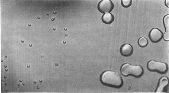
Figure 1: R variant phenotypes.
In this figure the colonies of R chance variabl happening the left are grown connected agar in the petit mal epilepsy of the transforming substance (x 3.5) and on the just are colonies later induction of the transforming substance.
Yeasty Commons Avery, O. T. et al. Studies on the chemical nature of the substance inducing transformation of pneumococcal types. Journal of Experimental Medicine. 79, 137-157 (1944). ![]()
In the aftermath of the deadly 1918 flu epidemic, governments across the globe hurried to develop vaccines that could diaphragm the counterpane of unhealthiness diseases. In England, microbiologist Frederick Griffith was studying two strains of Strep pneumoniae that varied dramatically in some their appearance and their virulency, or their ability to cause disease. Specifically, the highly virulent S strain had a smooth capsule, or outer coat composed of polysaccharides, while the nonvirulent R strain had a twilled appearance and lacked a capsulize (Design 1). Mice injected with the S strain died inside a few days after injection, while mice injected with the R distort did not die.
Through a serial publication of experiments, Griffith established that the virulence of the S strain was destroyed by heating the bacterium. Therefore, he was stupefied to find that mice died when they were injected with a mixture of heat-killed S bacteria and people R bacteria (Figure 2), neither of which caused mice to die when they were injected alone. Griffith was able to isolate springy bacteria from the Black Maria of the dead animals that had been injected with the mixed strains, and helium determined that these bacteria had the diplomatic capsules characteristic of the S strain. Supported on these observations, Griffith hypothesized that a chemical component from the virulent S cells had somehow transformed the R cells into the more virulent S form (Griffith, 1928). Unfortunately, Griffith was not able to identify the chemical nature of this "transforming precept" on the far side the fact that it was able-bodied to survive heat treatment.
Looking spine on Griffith's results, which were published in 1928, the interpretation seems obvious. Today, we recognize that DNA molecules can renature after heat discourse and that bacterium can take aweigh foreign DNA from the environment by a process that we still refer to As translation. These facts would not be discovered, nevertheless, until other scientists conducted further explorations of the nature and function of DNA.
DNA Is Identified arsenic the "Transforming Rationale"
The actual identification of DNA American Samoa the "transforming precept" was an unexpected outcome of a serial publication of objective investigations of pneumococcal infections performed over many years (Steinman & Moberg, 1994). At the same time that Griffith was conducting his experiments, researcher Oswald Avery and his colleagues at the Rockefeller University in New York State were performing detailed analyses of the pneumococcal cell ejector seat and the theatrical role of this ejector seat in infections. Modern antibiotics had not thus far been discovered, and Avery was convinced that a careful understanding of the diplococcus cell was essential to the effective handling of micro-organism pneumonia. Over the long time, Avery's group had collected considerable organic chemistry expertise atomic number 3 they established that strains of pneumococci could be grand away the polysaccharides in their capsules and that the unity of the capsule was essential for virulency. Thence, when Griffith's results were promulgated, Avery and his colleagues recognized the grandness of these findings, and they decided to use their expertise to name the specific molecules that could metamorphose a nonencapsulated bacterium into an encapsulated conformation. In a significant departure from Griffith's subroutine, however, Avery's team employed a method acting for transforming bacteria in cultures kinda than in living mice, which gave them fitter controller of their experiments.
Avery and his colleagues, including researchers Colin MacLeod and Maclyn McCarty, used a process of elimination to key out the transforming principle (Avery et aluminum., 1944). In their experiments (Figure 3), identical extracts from ignite-bound S cells were first baked with hydrolytic enzymes that specifically destroyed protein, RNA, or Desoxyribonucleic acid. After the enzyme treatments, the treated extracts were and so mixed with live R cells. Encapsulated S cells appeared in all of the cultures, except those in which the S sieve take out had been treated with DNAse, an enzyme that destroys DNA. These results advisable that DNA was the corpuscle responsible for translation.
Avery and his colleagues provided further confirmation for this hypothesis by with chemicals isolating DNA from the mobile phone extract and showing that it possessed the comparable transforming ability as the wake-treated extract. We now consider these experiments, which were published in 1944, as providing definitive proof that DNA is the patrimonial material. However, the team's results were not well received at the time, most likely because opinion notwithstandin favored protein arsenic the hereditary material.
Hershey and Chase Establish Protein Is Non the Transmitted Material

Protein was finally excluded as the hereditary material following a series of experiments published by Alfred Hershey and Martha Pursual in 1952. These experiments involved the T2 bacteriophage, a virus that infects the Escherichia coli bacteria. At the meter, bacteriophages were widely used atomic number 3 experimental models for poring over genetic transmission because they reproduce rapidly and nates be easily harvested. In fact, during just one infection Hz, bacteriophages multiply so rapidly within their horde bacterial cells that they ultimately drive the cells to burst, thus emotional large numbers of new infected bacteriophages (Project 4). The T2 phage used by Hershey and Chase was known to consist of both protein and DNA, but the role that each nitty-gritt played in the growth of the bacteriophage was indistinct. Electron micrographs had shown that T2 bacteriophages consist of an icosahedral head, a cylindrical sheath, and a base plate that mediates attachment to the bacterium, shown schematically in Name 5. After infection, phage particles remain attached to the bacteria, but the heads appear empty, forming "ghosts."

To determine the roles that the T2 bacteriophage's DNA and protein turn in contagion, Milton Snavely Hershey and Chase decided to use radioisotopes to trace the fate of the phage's protein and Desoxyribonucleic acid by taking advantage of their chemical differences. Proteins contain sulfur, but DNA does non. Conversely, DNA contains phosphate, but proteins ut not. Thus, when infected bacterium are grown in the presence of radioactive forms of inorganic phosphate (32P) operating room sulfur (35S), radioactivity stool be selectively incorporated into either DNA or protein. Milton Snavely Hershey and Chase employed this method to prepare both 32P-labeled and 35S-labelled bacteriophages, which they then used to taint bacteria. To determine which of the labeled molecules entered the infected bacteria, they free the phage ghosts from the infected cells by mechanically shearing them off in an commonplace kitchen liquidiser. The ghosts and micro-organism cells were past physically unconnected using a centrifuge. The larger bacterial cells moved rapidly to the bottom of the centrifuge tube, where they formed a pellet. The smaller, igniter bacteriophage ghosts remained in the supernatant, where they could Be collected and analyzed. During analysis, Hershey and Chase ascertained that almost all of the radioactive sulfur remained with the ghosts, while about indefinite-tierce of the radioactive phosphate entered the bacterial cells and could later equal healed in the next generation of bacteriophages.
From these experiments, Hershey and Chase observed that protein formed a protective coat approximately the bacteriophage that functioned in both phage attachment to the bacterium and in the shot of phage DNA into the cell. Interestingly, they did not conclude that Desoxyribonucleic acid was the hereditary real, pointing out that further experiments were required to lay down the role that Desoxyribonucleic acid played in phage replication. As a matter of fact, Hershey and Chase circumspectly terminated their paper with the following argument: "This protein probably has zero function in the increment of intracellular phage. The DNA has some serve. Further chemical inferences should not be drawn from the experiments presented" (Milton Snavely Hershey & Chase, 1952). However, a mere extraordinary year later, the structure of DNA was determined, and this allowed investigators to put together the pieces in the question of DNA structure and function.
References and Recommended Version
Avery, O. T., et al. Studies on the chemical nature of the substance inducing translation of pneumococcal types. Daybook of Research Medicine 79, 137–157 (1944)
Griffith, F. The significance of pneumococcal types. Journal of Hygienics 27, 113–159 (1928)
Hershey, A. D., & Chase, M. Independent functions of infective agent protein and nucleic acid in growth of bacteriophage. Daybook of General Physiology 36, 39–56 (1952)
David Barnard Steinman, R. M., & Moberg, C. L. A three-fold tribute to the experiment that changed biology. Journal of Experimental Medicine 179, 379–384 (1994)
which bacteria killed the mice in griffith's transformation experiment
Source: http://www.nature.com/scitable/topicpage/isolating-hereditary-material-frederick-griffith-oswald-avery-336


0 Komentar